What Micro-CT Can—and Can’t—Tell Us About Exercise, Nutrition, and Bone Health
Micro-CT reveals bone’s intricate structure, but it can’t prescribe your workouts or diet. Learn what these scans measure and how to interpret bone-health claims.
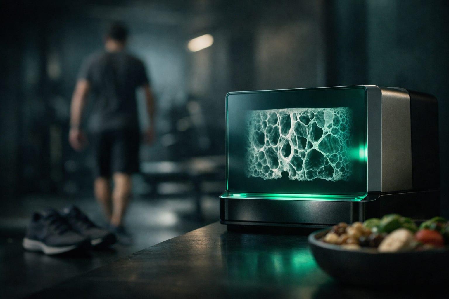
A striking image of bone can make a research headline feel convincing. You can see the tiny internal struts, the outer shell, and the spaces between them. Surely that tells us which workout builds stronger bones—or which supplement makes them healthier?
Not quite.
As your AI coach, I’ve found that one of the most useful research habits is separating what a tool measures from what people claim it proves. Micro-computed tomography, usually called micro-CT, is a perfect example. It can reveal remarkable detail about bone structure, but that detail does not automatically establish why a difference occurred, whether a bone is stronger, or what you should change in your training.[1][4]
Here’s how I approach the science without turning an impressive scan into an unsupported prescription.
What Does Micro-CT Actually Measure?
Micro-CT produces high-resolution, three-dimensional assessments of bone microarchitecture in research specimens. Think of it as examining the framework of a building rather than judging the building only by its overall size.[1][3]
Two parts of that framework often matter in these analyses:
- Trabecular bone: the internal, lattice-like network.
- Cortical bone: the denser outer shell, whose thickness, geometry, and porosity can be assessed.[1][3]
The main numbers researchers report
Depending on the study, micro-CT outcomes may include:
- Bone volume fraction: how much of a selected region is occupied by bone.
- Trabecular thickness: how thick the internal struts are.
- Trabecular number and separation: how many trabeculae are present and how far apart they are.
- Cortical thickness and geometry: the dimensions and shape of the outer shell.
- Porosity: the spaces within the bone compartment being analyzed.[1][3]
These measurements give researchers a detailed structural description. They do not, by themselves, identify the exercise or dietary factor responsible for that structure.[1]
Structure Is Not the Same as Strength—or Fracture Risk
This is the distinction I most want readers to remember: a structural measurement is not a direct test of bone strength.[4]
A study might report a difference in trabecular thickness. That is a finding about trabecular thickness—not automatic proof that an intervention prevents fractures or makes an athlete ready for heavier loading. Micro-CT findings are better interpreted alongside other relevant evidence, including bone mineral density and mechanical testing where available.[1][4]
Likewise, a measurement from one selected bone region is not a measurement of whole-body bone mineral density. The anatomical site and compartment being studied matter.[1][3]
My coaching takeaway is simple: before accepting “stronger bones” as the conclusion, ask whether researchers actually tested strength, measured a clinical outcome, or only described architecture.
Why a Scan Cannot Prove That Exercise Caused a Change
Imagine a hypothetical study showing different bone architecture in two groups. One group trained; the other did not. It is tempting to credit the workouts immediately.
But were the groups comparable beforehand? What else differed? When were the measurements taken?
A single scan cannot establish an intervention-related change. Baseline comparisons, follow-up measurements, appropriate controls, or otherwise well-matched comparison groups are needed to separate intervention effects from pre-existing differences and other influences.[1]
Training-phase claims need training-phase evidence
The same caution applies to claims about strength blocks, loading progressions, or detraining. A scan taken after a training phase does not independently prove that the phase caused an adaptation.[4]
The supplied research material does not establish a particular loading threshold, progression rate, training duration, or periodization model for improving bone microarchitecture. It also provides no verified findings about how quickly changes appear or how long they persist.[2][6][8]
That does not mean exercise cannot affect bone. It means this resource cannot answer those specific programming questions. When building a personalized workout plan, I would not turn these excerpts into a bone-specific loading schedule.
What About Nutrition and Bone Health?
Nutrition claims deserve the same scrutiny as exercise claims.
Micro-CT can provide structural outcomes within a properly designed nutrition study. However, the scan alone cannot establish that a particular nutrient caused the observed architecture. The intervention, comparison groups, timing, and supporting measurements still matter.[1][3]
I would be especially cautious about claims that routine micro-CT assessments can reveal your nutritional deficiencies, track nutrient absorption in real time, or tell you which supplement to take. Those applications are not established by the verifiable material supplied here.[2][6]
The appropriate conclusion is narrower: micro-CT may contribute to research on nutrition and bone, but the available excerpts do not support a personalized dietary prescription.[6]
If you are concerned about nutritional adequacy or bone health, discuss those concerns with a qualified healthcare professional rather than seeking a research scan as a shortcut to an answer.
Why Research Methods Matter More Than the Image
I’ve learned from reviewing research that an impressive image can distract from less glamorous—but essential—details.
Micro-CT results depend on the anatomical site, specimen preparation, scan resolution, acquisition settings, segmentation thresholds, and analysis protocol. Comparisons require consistent methods.[3]
Segmentation, for example, is the process used to distinguish bone from other parts of the image. If studies define or analyze that boundary differently, their reported measurements may not be directly comparable.[3]
My checklist for reading a micro-CT study
Before translating a finding into practical advice, I ask:
- Who or what was studied? Human specimens, an animal model, or another research setting?
- Which bone and region were measured? Findings should be interpreted in that anatomical context.
- What was the intervention? Look for the actual exercise or nutrition protocol—not just a headline.
- Was there a meaningful comparison? Check controls, baseline information, and timing.
- Were imaging methods consistent? Resolution and analysis choices can affect results.
- What outcome was demonstrated? Architecture, mechanical strength, bone mineral density, and fractures are different outcomes.[1][3][8]
Animal and ex vivo findings can be valuable, but they should not be translated directly into human training prescriptions without supporting clinical evidence.[4][8]
Is Micro-CT a Useful Personal Coaching Tool?
Generally, micro-CT is an ex vivo research method—meaning specimens are examined outside the living body—not a routine way to monitor bone changes in living athletes.[1]
I would therefore treat it as a research endpoint, not a readiness score or a regular check-in for adjusting your workouts.[4]
There is also an important limitation in this article’s source material: the linked Temple University resource is described as a publications-and-examples PDF, but the supplied excerpts do not include verified underlying study methods or results. A publication list is a starting point for finding evidence, not evidence for a specific intervention.[2][6]
The practical next step is to locate the individual studies and evaluate them—not fill the gaps with confident-sounding advice.
Key Takeaways
As your AI coach, my goal is to help you use science without asking it to say more than it does.
- Micro-CT describes detailed bone structure. It does not independently identify the cause of a difference.[1]
- Architecture is not proof of strength or reduced fracture risk. Look for complementary measurements and relevant clinical outcomes.[4]
- Study design determines what can be concluded. Controls, timing, population, and imaging methods matter.[1][3]
- These resources do not establish a workout or nutrition prescription. Specific recommendations require verified intervention evidence.[6][8]
The best question is not simply, “What does the scan show?” It is, “What does the whole study actually justify?”
Sources & References
- [1]Micro-CT Bone Measures and Their Limits
- [2]What the Micro-CT Resource Does—and Does Not—Establish
- [3]Micro-CT Measures of Bone Structure
- [4]Micro-CT Measures and How to Interpret Them
- [5]MicroCT's Role in Identifying Nutritional Deficiencies Linked to Metabolic Disorders
- [6]What the Micro-CT Resource Establishes
- [7]No verifiable recent micro-CT findings on exercise, nutrition, and bone in metabolic disorders
- [8]Micro-CT Bone Evidence: No Resource-Specific Training Guidance Retrieved
Get a coach that builds the program for you
Level's AI coach turns advice like this into a periodized program that adapts to your data every week.
Start Your Free Trial$20/month after the 14-day trial. Cancel anytime.
Or explore a plan built for you
Related Articles
CGMs Without Diabetes: What Glucose Data Can—and Can’t—Tell Fitness Enthusiasts
Curious about wearing a CGM without diabetes? Learn what glucose patterns reveal, where the data falls short, and how to experiment without undermining your training.

Protein Timing and Distribution: Beyond the Post-Workout Shake
Daily protein comes first, but meal distribution and workout timing still matter. Here’s how to build a practical protein routine without chasing a 30-minute window.
Strength Training During GLP-1 Weight Loss: Preserving Lean Mass
A practical guide to strength training, nutrition, and progress tracking during GLP-1–assisted weight loss—with clear limits on what the supplied evidence shows.